
 Q. No.
Match the following columns and select the correct option:
Column-I
Column-I
(a)
Eosinophils
(i)
Immune response
(b)
Basophils
(ii)
Phagocytosis
(c)
Neutrophils
(iii)
Release histaminase,
destructive enzymes
(d)
Lymphocytes
(iv)
Release granules containing histamine
(a)
(b)
(c)
(d)
1.
(iv)
(i)
(ii)
(iii)
2.
(i)
(ii)
(iv)
(iii)
3.
(ii)
(i)
(iii)
(iv)
4.
(iii)
(iv)
(ii)
(i)
destructive enzymes


Which of the following is associated with a decrease in cardiac output?
(1) Sympathetic nerves
(2) Parasympathetic neural signals
(3) Pneumotaxic center
(4) Adrenal medullary hormones
The QRS complex in a standard ECG represents:
1. Depolarisation of auricles
2. Depolarisation of ventricles
3. Repolarisation of ventricles
4. Repolarisation of auricles
Single circulation is characteristic of
1. Fish heart
2. Frog heart
3. Crocodilian heart
4. Marsupial heart
Three chamber heart of frog is not as efficient as four chambered human heart because :-
1. Oxygenated and deoxygenated blood mix up
2. Ventricle does not pump blood properly
3. It does not hold enough blood
4. Heart muscles are not strong
The decrease in the rate of heartbeat, speed of conduction of action potential and thereby the cardiac output is under the control of
1. Neural signals through the sympathetic nerves
2. Neural signals through the parasympathetic nerves
3. Adrenaline
4. ANS
Given below is the ECG of a normal human. Which of its components is correctly interpreted below?
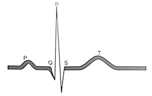
1. Peak P and Peak R together - Systolic and diastolic blood pressure
2. Peak P - Initiation of left atrial contraction only
3. Complex QRS - One complete pulse
4. Peak T - Initiation of total cardiac contraction
A special neural center that can moderate the cardiac function is located in
1. Cerebrum
2. Pons
3. Medulla oblongata
4. Cerebellum
Lymph ultimately release the absorbed substances into
1. Lymphatic capillaries
2. Bloodstream (veins)
3. Lymph nodes
4. Lymphatic duct
© 2024 GoodEd Technologies Pvt. Ltd.