
 Q. No.
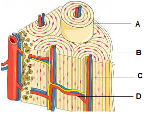
In the given diagram of ultrastructure of a compact bone, identify A, B, C and D:
1.
Osteon, Lamellae, Haversian canal, Volkmann’s canal respectively
2.
Lamellae, Osteon, Haversian canal, Volkmann’s canal respectively
3.
Lamellae, Osteon, Volkmann’s canal, Haversian canal respectively
4.
Osteon, Lamellae, Volkmann’s canal, Haversian canal respectively


In the given diagram of areolar tissue, the structure that secretes mediators of inflammation is labeled by the letter:

| 1. A | 2. B |
| 3. C | 4. D |
The type of connective tissue shown in A and B in the given diagram are respectively located in:
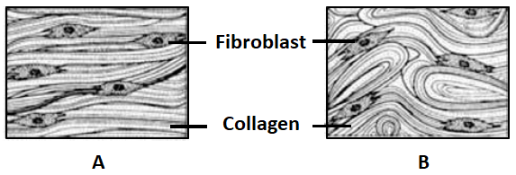
1. Tendon and Ligament
2. Ligament and Tendon
3. Tendon and Skin
4. Skin and Tendon


Identify the incorrect statement regarding the parts labeled in the following diagram:

1. A is the perimysium
2. B is the structural unit of skeletal muscle
3. C is epimysium
4. D is the endomysium
The epithelium shown in the figure is present in the lining of:
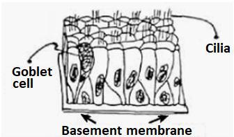
1. Fallopian tubes
2. Trachea
3. Ureter
4. Thyroid follicles
The type of exocrine gland seen in the lining of gastric mucosa is shown by the figure:
| 1. | 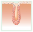 |
| 2. | 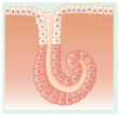 |
| 3. | 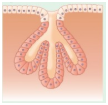 |
| 4. | 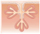 |


In the given diagram of a synovial joint, ligament and hyaline cartilage are represented respectively by the letters:

1. A and E
2. D and C
3. E and D
4. A and C
Based on the mode of secretion a gland shown in the following diagram will be called as:

1. Holocrine
2. Merocrine
3. Apocrine
4. Paracrine
The type of the neuron shown in the following diagram is seen in:

| 1. | Embryonic stages and Retina |
| 2. | Retina and Olfactory membrane |
| 3. | Retina and Dorsal root ganglion of spinal nerve |
| 4. | Olfactory membrane and Cerebellar peduncles |
In the given schematic diagram of ultrastructure of a myofibril, the functional unit of muscle contraction is shown by the letter:

| 1. | A | 2. | B |
| 3. | C | 4. | D |
© 2025 GoodEd Technologies Pvt. Ltd.