Ribosomes are the granular structures first observed under the electron microscope as dense particles by George Palade (1953). They are composed of ribonucleic acid (RNA) and proteins and are not surrounded by any membrane.
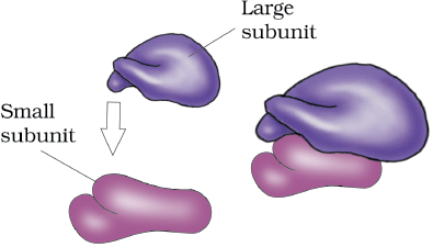
The eukaryotic ribosomes are 80S while the prokaryotic ribosomes are 70S. Each ribosome has two subunits, larger and smaller subunits (Fig 8.9). The two subunits of 80S ribosomes are 60S and 40S while that of 70S ribosomes are 50S and 30S. Here ‘S’ (Svedberg’s Unit) stands for the sedimentation coefficient; it is indirectly a measure of density and size. Both 70S and 80S ribosomes are composed of two subunits.
An elaborate network of filamentous proteinaceous structures consisting of microtubules, microfilaments and intermediate filaments present in the cytoplasm is collectively referred to as the cytoskeleton. The cytoskeleton in a cell are involved in many functions such as mechanical support, motility, maintenance of the shape of the cell.
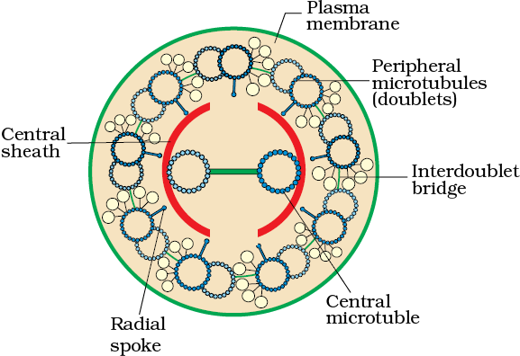
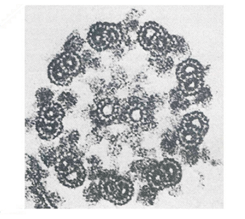
Cilia (sing.: cilium) and flagella (sing.: flagellum) are hair-like outgrowths of the cell membrane. Cilia are small structures which work like oars, causing the movement of either the cell or the surrounding fluid. Flagella are comparatively longer and responsible for cell movement. The prokaryotic bacteria also possess flagella but these are structurally different from that of the eukaryotic flagella.
© 2026 GoodEd Technologies Pvt. Ltd.

