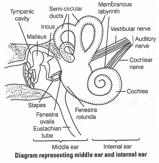
31. Explain the structure of middle and internal ear with the help of diagram.
Structure of Middle Ear
The middle ear contains three bones or ossicles-the malleus (hammer), incus (anvil) and stapes (stirr-up). These bones are attached to one another in a chain-like fashion. The malleus is attached to the tympanic membrane and the stapes is attached to the oval window (a membrane beneath the stapes) of cochlea. These three ossicles increase the efficiency of transmission of sound waves to the inner ear.
The middle ear also opens into the Eustachian tube, which connects with the pharynx and maintains the pressure between the middle ear and the outside atmosphere.
Structure of Internal Ear
The inner ear consists of a labyrinth of fluid-filled chambers within temporal bone of the skull. The labyrinth consists of two parts, i.e., the bony and membranous labyrinth. The bony labyrinth is a series of channels.
Inside these channels, membranous labyrinth lies, which is surrounded by a fluid called perilymph. The membranous labyrinth is filled with a fluid called endolymph. The coiled portion of the labyrinth is called cochlea.
The cochlea has two large canals-an upper vestibular canal (scala vestibuli) and a lower tympanic canal (scala typmani) - separated by a small cochlear duct (scala media). The vestibular and tympanic canals contain perilymph and the cochlear duct is filled with endolymph.
At the base of scale vestibuli, the wall of membranous labyrinth comes in contact with the fenestra ovalis, while at the lower end of scala tympani lies the fenestra rotunda.
© 2026 GoodEd Technologies Pvt. Ltd.

